The cerebellum, which stands for ‘little brain,’ is a hindbrain structure that controls balance, coordination, movement, and motor skills, and it is thought to be important in processing some types of memory.

In psychology, the cerebellum is often defined as a brain region responsible for coordinating and refining motor movements, ensuring balance and posture, and facilitating procedural learning.
While traditionally associated primarily with motor control, recent research has expanded our understanding of the cerebellum’s role, suggesting its involvement in cognitive processes, emotion regulation, attention, and even some aspects of language processing.
Thus, in a psychological context, the cerebellum is related to the physical coordination of movements and the coordination of thoughts and emotions.
Although the cerebellum only accounts for 10% of the overall brain mass, it contains over half of the nerve cells than the rest of the brain combined.
The cerebellum is also one of the few mammalian brain structures where adult neurogenesis (the development of new neurons) has been confirmed.

The cerebellum is an older part of the brain, also found in animals. According to scientists, it is even believed that the cerebellum was present in animals that existed before humans.
Where Is It Located?
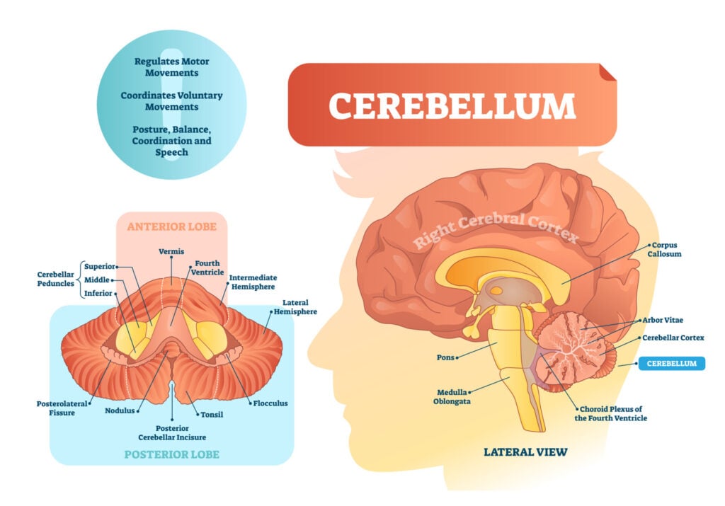
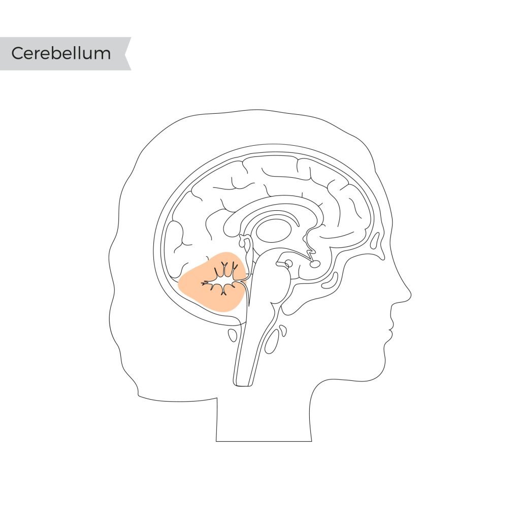
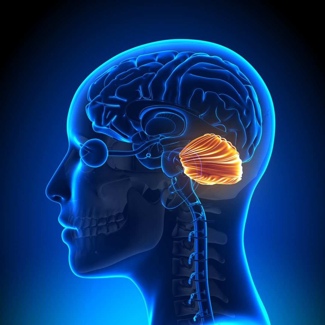
The cerebellum is located at the back of the brain, behind the brainstem, below the temporal and occipital lobes, and beneath the back of the cerebrum.
The cerebellum is also divided into two hemispheres, like the cerebral cortex. Unlike the cerebral hemispheres, each hemisphere of the cerebellum is associated with each side of the body.

Structure
The cerebellum consists of the cerebellar cortex, the outer layer, containing folder brain tissue, filled with most of the cerebellum’s neurons. It plays a key role in processing and integrating information sent to the cerebellum.
There is also a fluid-filled ventricle and cerebellar nuclei, which is the innermost part, containing neurons that communicate information from the cerebellum to other areas of the brain.
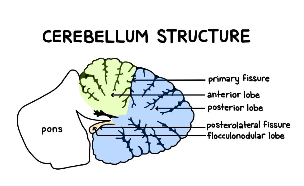
Within the cerebellum, there are thought to be three anatomical lobes, which are divided by two fissures (large furrows)– the primary fissure and the posterolateral fissure:
- Anterior lobe (anterior meaning ‘to the front’): Primarily involved in the coordination of limb movements.
- Posterior lobe (posterior meaning ‘to the back’): The largest part and plays a significant role in planning, initiating, and timing movements.
- Flocculonodular lobe: The oldest part of the brain in terms of evolution. This part of the cerebellum is responsible for balance and spatial attention, as well as receiving visual input.

The cerebellum can also be divided into three functional areas:
- Cerebrocerebellum – this is the largest area of the cerebellum, responsible for planning movements and motor learning. It also works to regulate the coordination of muscle activation as well as eye movements.
- Spinocerebellum – this area functions in regulating body movements by allowing for error corrections.
- Vestibulocerebellum – this area is involved in controlling balance and flexes of the eyes.
Functions
The cerebellum, located at the base of the brain, is responsible for coordinating voluntary movements, maintaining posture, balance, and equilibrium, as well as refining motor movements to be smooth and precise. It also plays a role in some cognitive functions, such as attention and language processing.
The cerebellum’s main role is to monitor and regulate motor behavior without any need for conscious awareness.
It was once believed that the only function of the cerebellum was to coordinate movements. We now, however, understand that the cerebellum plays a much bigger role in a variety of functions and communicates signals to other areas of the brain.
Below is a list of some of the associated functions of the cerebellum:
- Coordination of voluntary movement.
- Balance.
- Posture.
- Motor-learning.
- Sequence learning.
- Reflex memory.
- Mental function.
- Emotional processing.
The cerebellum receives sensory information, especially regarding the body’s position, so it knows what each body part is doing. Signals can be received from the brain stem, spinal cord, and cerebrum, to coordinate and control movement.
It receives information from the frontal lobes of the brain, so it knows what movements the frontal lobes intend to make. Although the cerebellum does not initiate movement, it does help to organize the movements to ensure it is a fluid and coordinated action.
On their own, the frontal lobes would produce jerky, uncoordinated, and inaccurate movements, therefore the cerebellum plays an important part in regulating this. Fundamentally, the cerebellum organizes all muscle group actions, including eye movements.
The cerebellum also aids in balance and posture. It monitors information regarding balance and posture to ensure that when we are standing or walking, we are not falling down and we are able to keep ourselves steady.
In terms of motor learning, the cerebellum is vital when learning a new skill. For instance, if someone were learning to ride a bike for the first time, you may expect they would typically start off making mistakes and falling off the bike.
The cerebellum helps to fine-tune the motor skills required to ride a bike until they get to a point where the action can be completed seamlessly and almost automatically.
Damage
Cerebellar damage results in the breakdown and destruction of nerve cells which can have long-last effects. A person who has damage to their cerebellum may experience some of the following symptoms:
- Walking unsteadily
- Tremors – involuntary rhythmic contractions
- Vertigo – which can also lead to swaying, nausea, and headaches.
- Slurred speech
- Inaccurate or jerky movements
- Cognitive impairment – this can affect memory, learning, and thinking.
- Dystonia – involuntary contractions of muscles, which can result in muscles being held in painful positions as a result
- Clumsiness – may give someone the appearance of being intoxicated
- Less refined motor-skills
- Ataxia – loss of control of voluntary movement
Drinking alcohol has an immediate and temporary effect on the cerebellum as the body’s coordination and movements become clumsy. Someone who is intoxicated with alcohol may not be able to walk in a straight line and lose their balance.
Although these symptoms are temporary, repeated alcohol misuse, becoming an alcohol use disorder, can have long-lasting impacts on the cerebellum and lead to these symptoms being more long-lasting.
Other causes of damage to the cerebellum can come from injury to the head, such as falling backward and hitting the back of the head where the cerebellum lies. Brain tumors and infections in the brain can also cause long-lasting damage to the cerebellum.
Damage could also occur through medical issues such as having Parkinson’s disease, multiple sclerosis, and experiencing a stroke. Similarly, lead or mercury poisoning and the overuse of certain medications (e.g., benzodiazepines) can also cause lasting damage to cerebellar function.
Research Studies
- Schmahmann & Sherman (1998) investigated behavioral changes in patients who had diseases confined to the cerebellum. They found that these patients had impairments in executive functions such as planning, working memory and abstract reasoning.
- They also found difficulties in visual and spatial organization and some language deficits.
- They concluded that the cerebellum plays a big part in the areas of language and cognition.
- Brown et al. (2005) examined the neural basis of dance and they found evidence of cerebellar activity whilst synchronizing timing and movement to musical rhythm.
- Ponti, Peretto & Bonfanti (2008) investigated the cerebellums of adult rabbits and found evidence of neurogenesis (the development of new nerve cells).
- Jimsheleishvili & Dididze (2019) proposed the cerebellum is involved in processing language and mood, attention, the fear response as well as the pleasure and reward response.
- Stoodley & Schmahmann (2009) conducted neuroimaging studies of the cerebellum. They found that various parts of the cerebellum become activated during tasks involving movement and touch, spatial memory, emotional processing, and verbal memory.
- They also found that language was associated with the right-hand side of the cerebellum, whereas the left side of the cerebellum was associated with spatial awareness.
- Leggio et al. (2008) found that there were cognitive sequencing impairments in patients with damage to their cerebellum.
- Appollonio et al., (1993) found that the ability to produce words fluently, as well as problem-solving, are impaired in patients whose cerebellum has been damaged.
- Caulfield & Servatius (2013) propose that the cerebellum is involved in those with anxiety disorders. They suggest that because avoidance (which is a trait of anxiety disorders) is a learned process that has been reinforced over time, that this has been learned through the aid of the cerebellum.
- They found that lesioning the cerebellum in rats prevented them from using the avoidance response to anxiety-provoking situations.
- Impairment to the cerebellum has been reported in those with anxiety disorders and may be linked to increased arousal present in posttraumatic stress disorder (PTSD) and generalized anxiety disorder (Abadie et al.,1999).
- Similarly, cerebellar hyperactivity has been shown to correlate positively with increase blood pressure and heart rate, a possible reason for the cerebellum’s role in anxiety disorders (Critchley et al., 2000.)
- Stoodley (2016) suggested that the cerebellar dysfunction is found in individuals with developmental disorders such as Autism, attention deficit hyperactivity disorder (ADHD) and developmental dyslexia.
- They also suggested that early damage to the cerebellum has been shown to be associated with poorer outcomes in comparison to damage in adulthood. This implies that protecting the cerebellum is vital during childhood development.
- Gowen & Miall (2007) aimed to test whether there was any evidence of cerebellar involvement in motor dysfunctions associated with neuropsychiatric disorders.
- There was some evidence found that Autism, schizophrenia, and dyslexia is linked to the cerebellum, although they are not restricted to this brain area.
- Brain imaging studies have found that there are reduced sizes of the cerebellum in patients who are diagnosed with schizophrenia (Nopoulos et al., 1999).
- There is also evidence from neuroimaging that patients with schizophrenia have less blood flow to the cerebellar cortex during the performance of cognitive tasks, such as attention and tasks which involve using short-term and working memory (Crespo-Facorro et al., 2007).
Summary
There is growing research and data to suggest that the cerebellum plays a role, not only in controlling balance and voluntary movement, but also in the control of cognitive and emotional processes.
There is also evidence through neuroimaging studies to suggest the cerebellum’s involvement in neurological disorders.
In order to preserve the health of the cerebellum, limiting or stopping smoking and drinking alcohol is a suggestion. This is because they both contribute to raising blood pressure which could ultimately lead to a stroke.
Exercising more and eating a healthy diet both help lower blood pressure, and thus, the risk of a stroke, so this is encouraged to protect the cerebellum.
Finally, protecting the head in general, such as wearing helmets when cycling, wearing seatbelts in the car, and taking care to prevent falls in the home, can limit the risk of damage to this area of the brain.
References
Abadie, P., Boulenger, J. P., Benali, K., Barre, L., Zarifian, E., & Baron, J. C. (1999). Relationships between trait and state anxiety and the central benzodiazepine receptor: a PET study. European Journal of Neuroscience, 11 (4), 1470-1478.
Appollonio, I. M., Grafman, J., Schwartz, V., Massaquoi, S., & Hallett, M. (1993). Memory in patients with cerebellar degeneration. Neurology, 43 (8), 1536-1536.
Brown, S., Martinez, M. J., & Parsons, L. M. (2006). The neural basis of human dance. Cerebral cortex, 16(8), 1157-1167.
Caulfield, M. D., & Servatius, R. J. (2013). Focusing on the possible role of the cerebellum in anxiety disorders. New Insights into Anxiety Disorders (Durbano F, Ed.). InTech, Rijeka, HR, 41-70.
Crespo-Facorro, B., Barbadillo, L., Pelayo-Terán, J. M., & Rodríguez-Sánchez, J. M. (2007). Neuropsychological functioning and brain structure in schizophrenia. International Review of Psychiatry, 19 (4), 325-336.
Critchley, H. D., Corfield, D. R., Chandler, M. P., Mathias, C. J., & Dolan, R. J. (2000). Cerebral correlates of autonomic cardiovascular arousal: a functional neuroimaging investigation in humans. The Journal of physiology, 523(1), 259-270.
Gowen, E., & Miall, R. C. (2007). The cerebellum and motor dysfunction in neuropsychiatric disorders. The Cerebellum, 6(3), 268-279.
Jimsheleishvili, S., & Dididze, M. (2019). Neuroanatomy, cerebellum. StatPearls.
Leggio, M. G., Tedesco, A. M., Chiricozzi, F. R., Clausi, S., Orsini, A., & Molinari, M. (2008). Cognitive sequencing impairment in patients with focal or atrophic cerebellar damage. Brain, 131 (5), 1332-1343.
Nopoulos, P. C., Ceilley, J. W., Gailis, E. A., & Andreasen, N. C. (1999). An MRI study of cerebellar vermis morphology in patients with schizophrenia: evidence in support of the cognitive dysmetria concept. Biological psychiatry, 46 (5), 703-711.
Phillips, J. R., Hewedi, D. H., Eissa, A. M., & Moustafa, A. A. (2015). The cerebellum and psychiatric disorders. Frontiers in public health, 3, 66.
Ponti, G., Peretto, P., & Bonfanti, L. (2008). Genesis of neuronal and glial progenitors in the cerebellar cortex of peripuberal and adult rabbits. PLoS One, 3 (6), e2366.
Schmahmann, J. D., & Sherman, J. C. (1998). The cerebellar cognitive affective syndrome. Brain: a journal of neurology, 121(4), 561-579.
Stoodley, C. J. (2016). The cerebellum and neurodevelopmental disorders. The Cerebellum, 15 (1), 34-37.
Stoodley, C. J., & Schmahmann, J. D. (2009). Functional topography in the human cerebellum: a meta-analysis of neuroimaging studies. Neuroimage, 44(2), 489-501.

